2. Patty Infiltration Of The Liver
Description
This section is from the book "A Manual Of Pathology", by Joseph Coats, Lewis K. Sutherland. Also available from Amazon: A Manual Of Pathology.
2. Patty Infiltration Of The Liver
Fat is often found in large quantities in the liver in cases where, in the subcutaneous tissue or elsewhere, there is an actual deficiency of it. The fat in the liver is in the peripheral parts of the lobules, and from this it is to be inferred that it has been brought by the portal blood, and that it is a store fat. This fatty infiltration occurs most frequently in phthisis pulmonalis.
Its accumulation in diseases such as phthisis may, in part, be accounted for by supposing that the fat which is normally used for the formation of the fatty acids and the cholestearine of the bile is not so used, and is therefore stored in the hepatic cells. It is known that the secretion of bile is greatly diminished in such cases, and that the bile is watery. In that case the fatty infiltration here would, like that in muscle, be due to diminished activity of the organ. Another view, and one having some appearance of probability, is based on the theory that one of the functions of the liver is to prepare fat for oxidation. Naumann (Reichert and Du Bois Raymond's Archiv, 1871, p. 41) has shown that the liver fat is much more oxidizable than ordinary fat, and that in the vertebrata the size of the liver is in inverse proportion to the activity of the respiration, being largest in fishes and smallest in birds. It is therefore suggested that in phthisis and cachectic diseases the liver may produce an excess of easily oxidizable fat and store it up ready for use. Hence, perhaps, the utility of liver oils in cases of phthisis.
We have seen that in fatty degeneration the fat appears in the form of fine granules or drops, and that as these increase they remain isolated. In fatty infiltration there are, of course, first fine fat drops, but as more fat is added the drops grow in size. In the case of the conversion of connective tissue into adipose tissue, there is a single fat drop in each cell, as in Fig. 46. In the case of fatty liver the fat drops are of various sizes (see Fig. 47), but, as a rule, much larger than in fatty degeneration. The size is by no means an absolute criterion, but it is an important practical indication. It is only in the liver that there can be much difficulty in distinguishing between fatty degeneration and fatty infiltration, and here the fact that in the latter the fat is deposited at first in the cells at the peripheral parts of the lobules, and continues more abundant there, is sufficiently distinctive. There is one exception to this in the case of the fatty liver of alcoholism. Here a fatty infiltration occurs which is diffused throughout the lobules.
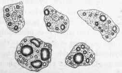
Fig. 47. - Fatty infiltration of the liver as seen in the fresh state. Isolated hepatic cells with drops of fat of various sizes, x 350.
Continue to:
My Books

