Conducting Tissue. Part 4
Description
This section is from the "Histology of Medicinal Plants" book, by William Mansfield. Also see Amazon: Histology of Medicinal Plants.
Conducting Tissue. Part 4
Structure Of Cells
The structure of the individual cells forming the medullary rays differs greatly in different plants, but is more or less constant in structure in a given species.
The medullary rays of the wood usually have strongly pitted side and end walls, while the medullary rays of most barks are not at all, or only slightly, pitted. In most plants the cells are of nearly uniform size. Frequently, however, the cells vary in size in a given ray, as shown in the cross-section of kava-kava.
Arrangement Of The Cells In A Ray
The union of any two cells in a ray is also of importance. In quassia the medullary ray cells have oblique end walls, so that on cross-section the line of union between two cells is an oblique wall. In most plants the medullary ray cells have blunt or square or oblique end walls, so that the line of union is a straight line.
In most plants the cells are much longer than broad, but the cells of sassafras bark are nearly as broad as long.
The walls of the cortical medullary ray cells and the medullary rays of most roots and stems of herbs are composed of cellulose; while the walls of medullary ray cells occurring in woods are frequently lignified.
There is a great variation in the character of the cell contents of medullary rays. In white pine bark (Plate 48, Fig. B1) are deposits of tannin; in quassia wood, starch; in canella alba, rosette crystals of calcium oxalate, etc.
Latex Tubes
Living latex tubes, like sieve tubes, have a layer of protoplasm lining the walls, and, in addition, have numerous nuclei. In drug plants the nuclei are not distinguishable, but the protoplasm is always clearly discernible.
Latex tubes function both as storage and as conducting cells. They, like the sieve tubes, contain proteid substances chiefly, yet frequently starch is found. The cells bordering the latex tubes absorb from them, as needed, the soluble food material. While our knowledge concerning the function of latex in some plants is meagre, still in other plants it is practically certain that the latex is composed of nutritive substances which are utilized by the plant as food. In certain other plants the latex appears to be used as a means of resisting insect attacks and as a protection against injury.
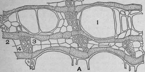
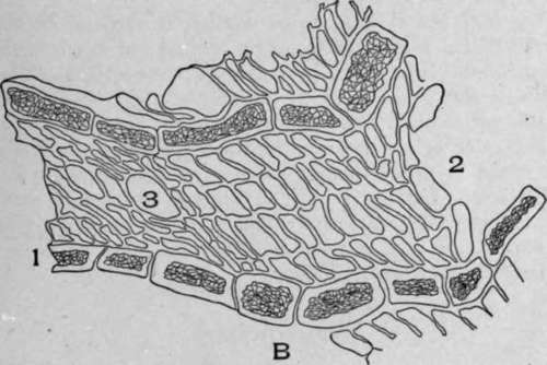
Plate 48. A. Cross-section of kava-kava root {Piper methysticum, Forst., f.).
1. Unequal diameter medullary ray cells.
2. Vessels.
3. Wood parenchyma.
4. Wood fibres.
B. Cross-section of white pine bark (Pinus strobus, L.).
1. Wavy medullary rays with tannin.
2. Parenchyma cells.
3. Sieve cells.
There are two types of latex tubes common to plants, namely, latex cells and latex vessels. Latex tubes developing from a single cell do not differ materially from a latex tube originating from the fusion of several cells. In each case the latex tube branches to such an extent that it bears no resemblance to ordinary cells. It would seem that the ultimate branches are formed and develop in much the same manner as root hairs - that is, by a growing tip of the branch. A mature plant may therefore have latex tubes with almost numberless branches (Plate 50, Fig. 1) and be of very great length.
The branches of latex tubes develop in such an irregular manner that it is possible to obtain a cross and a longitudinal section of the latex tubes by making a cross-section of stem. Such a section is shown in the drawing of the cross-section of the rhizome of black Indian hemp (Plate 49, Fig. B).
The color of the latex in medicinal plants varies from a gray white in papaw (carica papaya), aromatic sumac, black Indian hemp, and bitter root, to white in the opium poppy, light orange in celandine, and deep orange in bloodroot (Plate 50, Fig. 2). In each of these cases it is the latex which yields the important medicinal products.
Parenchyma
The larger amount of plant tissue is composed of parenchyma cells. These cells vary from square to oblong, or they may be irregular and branched. The end walls are square or blunt, and the wall is composed of cellulose, with the exception of the wood parenchyma, which has lignified walls.
There are seven characteristic types of parenchyma cells: (1) cortical parenchyma, (2) pith parenchyma, (3) wood parenchyma, (4) leaf parenchyma, (5) aquatic plant parenchyma, (6) endosperm parenchyma, (7) phloem parenchyma.
Parenchyma cells, cortical, pith, aquatic plant, leaf, flower, and endosperm, conduct in all directions - upward, downward, and laterally. The direction of conduction depends upon the needs of the different cells forming the plant. The fluids pass from the cell with an abundance of cell sap to the cell with less cell sap. In this wall all cells are provided with food.
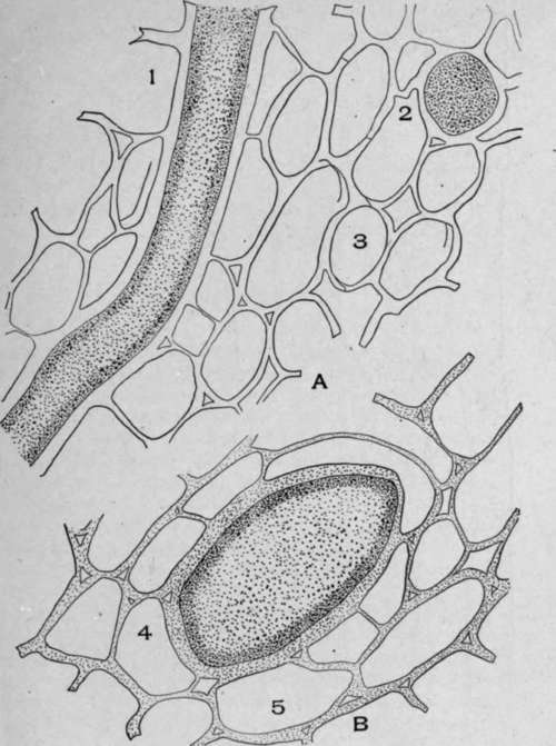
Plate 49. A. Cross-section of black Indian hemp (Apocynum cannabinum, L.).
1. Longitudinal section of a latex tube.
2. Cross-section of latex tube.
3. Parenchyma.
B. Cross-section of a part of black Indian hemp root.
4. Cross-section of a large latex tube.
5. Parenchyma.
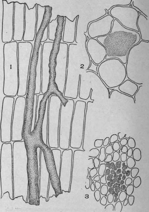
Plate 50. Latex Vessels.
1. Radial-longitudinal section of dandelion root (Taraxacum officinale, Weber).
2. Cross-section of sanguinaria root (Sanguinaria canadensis, L.).
3. Cross-section of dandelion root.
Parenchyma cells conduct water absorbed by the roots and soluble carbohydrate material chiefly.
The walls of all the different types of parenchyma cells are composed of cellulose with the exception of the wood parenchyma cells, the walls of which are lignified. The end walls of non-branched parenchyma cells and the cell terminations of branched cells are very blunt.
Continue to:
My Books

