Chapter 12, Note 1. Lipids and Cytolytic Activity of Sera (245, 246)
Description
This section is from the book "Research In Physiopathology As Basis Of Guided Chemotherapy With Special Application To Cancer", by Emanuel Revici. Also available from amazon: Research In Physiopathology
Chapter 12, Note 1. Lipids and Cytolytic Activity of Sera (245, 246)
The fact that cancer cells are found circulating in the blood without inducing metastases throughout the body, as might be expected, has made various workers investigate the means by which the organism rids itself of these cells and which might represent part of a general process through which the organism could fight cancer. Research to confirm the capacity of blood sera for influencing the lysis of cancer cells has been carried on in our laboratories by Robert Willheim and co workers. Sera of both normal individuals and cancer patients were found to induce lysis of cancer cells in vitro. Ehrlich, Krebs and Sa 180 ascites in mice were used, as well as cells obtained from solid tumors with encephaloid character. Tumors obtained from biopsies or autopsies of human beings also were used. Lytic activity was determined through the dilution of sera which later was mixed with a suspension of a known number of cells. After incubation for one hour
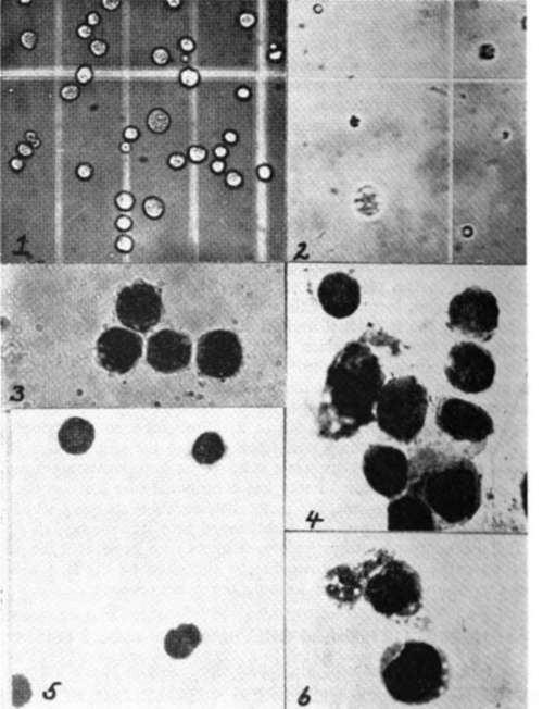
Fig. 278. Lytic action of human serum upon Sa 180 ascites cells.
(1) Normal SA 180 Cells X 300, not stained.
(2) Lytic effect of Serum on SA 180 X 300, not stained.
(3) Normal SA 180 Cells X 1000. Giemsa stain.
(4) Lytic effect of Serum on SA 180 Cells X 1000, Giemsa stain.
(5) Normal Krebs Cells X 1000. Giemsa stain.
(6) Lytic effect of Serum on Krebs Cells X 1000, Giemsa stain.
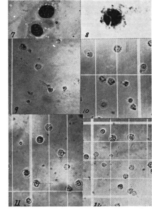
Fig. 279. Continuation.
(7) Normal SA 180 Cells X 1800. Giemsa stain.
(8) Carcinolytic effect of Serum on SA cells X 1800, Giemsa stain.
(9) Normal Lysis of SA 180 Cells as control in Polysaccharides Inhibition Studies on Carcinolysis X 300, not stained.
(10) Effect on Carcinolysis by the Polysaccharide Dextran X 300, not stained.
(11) Effect of Levan on Carcinolysis. Note the clearly distinct outline of unaffected cells X 300, not stained.
(12) Effect of Mucin on Carcinolysis X 300, not stained (evidence of inhibited Lysis.
at 37°C the product was examined and the cells counted in an adequate chamber. (Fig. 278) Relatively big variations were seen in different individuals, the results being negative for 1:1 dilution in some cases and positive in 1:32 and even higher for other individuals. No correlation could be established between this lytic power and the clinical condition of an individual.
With R. Willheim and M. Auber (247) we showed that the addition of a suspension of unsaturated fatty acids is able to induce such a lytic property in sera without this capacity, while the addition of insaponifiable fractions or of cholesterol inhibited lytic activity. With R. Willheim and M. Auber we (248) have investigated the problem of the correlation between the lytic activity and the structure of different fatty acids and their sodium soaps. The higher members of the fatty acid series have shown intense lytic activity. It could be shown that the lipoidic character of the fatty acids increases their lytic capacity. The value of the lipoidic character of the fatty acids used has appeared when members with the same number of carbons but having an hydroxyl or carboxyl in their molecule, were tested. With disappearance of lipoidic character, lytic property disappeared. The research covered not only cancer cells but also liver, red blood cells and lymphocytes. It could be seen that a correlation exists between the rapid growth character of these cells and the capacity of the fatty acids to attack them. It is interesting to note that cancer cells treated in vitro with fatty acids having lytic activity no longer produced cancerous growths when transplanted to animals. (249)
Continue to:
My Books

