Cytologic Examination Of The Urinary Sediment
Description
This section is from the book "Early Detection And Diagnosis Of Cancer", by Walter E. O'Donnell. Also available from Amazon: Early Detection And Diagnosis Of Cancer.
Cytologic Examination Of The Urinary Sediment
Collection of urine for cytologic study is something that can be done on the first visit to the office.
It is particularly important when the renal lesion is thought to arise in the kidney pelvis rather than in the parenchyma, since in the former location cells are particularly likely to exfoliate and produce a positive specimen.
Technique of collecting urine for cytologic study (Fig. 78)
1. Approximately 50 ml. of a catheterized specimen of urine is necessary for all female patients and is preferable for male patients. However, voided urine from the male may be used.
2. The specimen must be mixed with an equal amount of 50% ethyl alcohol.
3. Specimens should be sent to the cytology laboratory with complete clinical information.
4. If the specimen is to be mailed, the bottle should be tightly covered and inserted in mailing carton. If delay is unavoidable or if the specimens must be kept overnight, place them in a refrigerator.

Obtain approximately 50 ml. of catheterized urine.

Mix the specimen with equal amount of 50% ethyl alcohol.
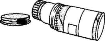
Cover tho specimen bottle tightly, anal place it in a mailing container clearly labeled and identified.
Fig. 78. Technique of collecting urine for cytologic study.
Results tend to vary rather widely, but in general a positive cytology can be expected:
1. In pelvic lesions: over 80% of cases
2. In parenchymal lesions: 10 to 15% of cases
If there is an obstruction to urinary flow from the renal pelvis to the ureter and bladder, the chances of obtaining a positive cytology are greatly diminished.
It must be remembered that parenchymal tumors are by far the more common and usually will not yield a positive urine cytology.
Hematology
The hemoglobin and/or hematocrit determination plus the white blood count should of course be included in the work-up of any kidney cancer suspect since they may give evidence of the following:
1. Anemia (most common)
2. Polycythemia
These findings, if present-and they frequently are (not simultaneously, however)-do not necessarily indicate an unfavorable prognosis.
Such abnormalities may disappear following removal of the tumor.
X-Ray Film Of The Chest
X-ray film of the chest should of course be included in the work-up of any cancer suspect.
In the case of possible renal cancer it is important to rule out the presence of pulmonary metastases, which would greatly influence the subsequent management of the case.
Cystoscopy And Retrograde Pyelography
If, after the preceding steps have been followed, there persists any suspicion of a renal tumor, cystoscopy and retrograde pyelography (Fig. 77) must be carried out and possibly followed by the more extensive radiographic procedures previously listed. Specialized training and equipment are required for these techniques.
Surgical exploration
No matter what the results of the foregoing studies are, if there remains persisting suspicion of renal cancer, surgical exploration may be required.
It is virtually impossible to be certain whether a given unilateral renal lesion, identified by physical examination, x-ray studies, etc., is benign or malignant without resorting to exploration.
Under these circumstances surgical exploration is usually advisable.
Continue to:
My Books

