Karyokinesis
Description
This section is from the book "A Manual Of Pathology", by Guthrie McConnell. Also available from Amazon: A Manual Of Pathology.
Karyokinesis
In karyokinesis, or indirect division, the cell goes through a very complicated course of changes of the various elements, probably the result of definite metabolic processes.
The changes can best be considered under four headings:
1. The Prophase
The centrosome increases in size, passes from the nucleus into the cytoplasm, and divides into two.
Surrounding each centrosome is a mass of fine radiating lines known as the amphiaster. The rays extending from one centrosome to another are arranged in spindle form, the centrosomes being situated at the apices of the spindles. These achromatin rays form the nuclear spindle.
The nucleus has been enlarging and the chromatin increasing, its particles uniting to form a long fuzzy thread. These fibrils become tangled and convoluted and form the close skein. The fibrils become thicker, less convoluted, and arrange themselves in irregular loops, forming the loose skein. The chromatin now stains much more deeply than normally. These loops finally separate at their peripheral ends and form the chromosomes, V-shaped fibrils with their closed ends arranged in a clear space known as the polar field.
During the formation of the skeins the nucleolus and the nuclear membrane disappear and the chromatin fibrils lie in the cell protoplasm.
The chromosomes are always present in the same number in the same species, varying from 2 to 50 in various animals; in man being 24.
The arrangement of the fibrils about the polar field constitutes the mother star or monaster.
2. The Metaphase
Each of the chromosomes undergoes a longitudinal division into two. These filaments, with the closed end advancing, begin to separate, moving toward their respective poles or centrosomes.

Fig. 16. - Nuclear Changes in Karyokinesis (Hatschek).
a, Nucleus of spermatoblast of Salamandra maculata, with chromatin threads forming the first suggestion of a coil; b, close coil with disappearance of the fuzzy aspect and longitudinal cleavage of the threads.

Fig. 17. - Diagrammatic Appearance of the Relation of the Chromosomes to the Centrosomes and Primitive Nuclear Spindle (Flemming).

Fig. 18. - Diagrammatic Representation of the Nuclear Spindle and of the Arrangementofthe Double Chromosomes in an Equatorial Plane Preparatory to Separation. This Stage is Called the Mother Star (Flemming).
3. The Anaphase
The Anaphase begins with the migration of the chromosomes. As they move toward the opposite poles the free ends constitute the equatorial plate. Connecting the ends are fine threads of achromatin known as the connecting filaments. The chromosomes collect at the opposite ends and form the daughter stars or Masters. As this occurs there is the beginning of a constriction of the protoplasm.

Fig. 19. - Diagrammatic Representation of the Separation of the Chromosomes, which are Attached toward Opposite Poles of the Nuclear Spindle, about which They Gather to Form the "Daughter Stars" (Flemming).
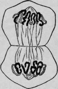
Fig. 20. - Segmentation of the Cytoplasm, and the Chromosomes Equally Divided, about to Form New Nuclei in the New Cells (Flemming).
4. The Telophase
The constriction continues until the original cell has been completely divided and two new ones formed. The chromosomes now undergo in reverse order the phases that have been described: the loose skein, the close skein, the reappearance of the nuclear membrane and of the nucleolus, with finally the stage of rest. To summarize, the changes are as follows: Resting mother nucleus. Prophase.
Migration and division of the centrosome with increase of chromatin. Close skein.
Disappearance of the nuclear membrane. Disappearance of the nucleolus. Loose skein.
Separation of the skein into chromosomes. Appearance of the polar field. Rearrangement of the chromosomes around polar field. Monaster, or mother star. Appearance of the nuclear spindle. Metaphase.
Longitudinal division of the chromosomes. Anaphase.
Migration of the divided chromosomes to opposite ends of the cell. Formation of the equatorial plate. Diaster or daughter star. Telophase.
Constriction of the protoplasm.
Daughter skeins undergoing in reverse order the above changes. The stage of rest. In some instances, instead of the cytoplasm dividing when cleavage of the nucleus is completed, it remains unchanged. This may go on until there are many nuclei, imbedded within a single mass of cytoplasm. Such formations are known as giant cells, and may be the result of division under unfavorable circumstances.
There may be the formation of more than two centrosomes with a resulting multipolar cell. The equatorial segments may split up more than once and the daughter cells may divide secondarily.
Continue to:
My Books

