1. Taenia Solium
Description
This section is from the book "A Manual Of Pathology", by Joseph Coats, Lewis K. Sutherland. Also available from Amazon: A Manual Of Pathology.
1. Taenia Solium
This form is of very common occurrence in this country, but that taken next is probably as frequent, if not more so. The strobilus or mature worm occurs in the alimentary canal, and the head is usually situated in the duodenum or upper part of the jejunum, while the rest of the animal extends downwards in the canal, attaining on an average a length of from ten to twelve feet. As already mentioned, this, like other tape-worms, has no alimentary canal, and supports itself by imbibition of nutritious material from the intestine.
The head of the worm, which is represented in Fig. 172 is about the size of a pin's head, and of a generally rounded form. In front it is prolonged so as to form a proboscis or rostellum, which is surrounded by a circle of twenty-six hooklets, which are alternately larger and smaller (see Fig. 179). The wide part of the head has four large sucking discs. On the head follows a narrow neck, which is so thin that it readily breaks when the worm is handled, rendering it difficult to obtain the small head. The proper neck is about half an inch in length, and it gradually merges in the anterior part of the body, in which fine transverse lines begin to appear as the first indications of the formation of segments. On passing down, the worm increases in breadth, while the segments elongate and become more completely divided. At first the segments or proglottides are homogeneous in appearance, but by and by the sexual apparatus begins to appear.
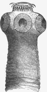
Fig. 172. - Head of T. solium X 45. (Leuckart).
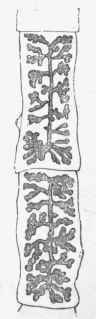
Fig. 173. - Two ripe proglottides of T. solium, With branches of uterus shown. x 2. (Leuckart).
The total number of segments in a worm ten feet in length is about 800. The sexual apparatus begins to appear about the 200th segment from the front, and is mature about the 450th; it consists of the male and female organs, which are present in each segment. In the fully matured segment the ova are visible, and when a proglottis is dried on a glass slide they indicate the form of the uterus, which in this tapeworm consists of a central stem and ramifying lateral branches to the number of seven to ten (see Fig. 173).
The male organs consist of a large number of vesicles scattered throughout the segment, as shown in Fig. 174, but more abundant anteriorly, as the female organs occupy the space behind. The vesicles are connected with fine seminal tubules, which are difficult to make out, and are shown in the figure as fine branching lines. These end in a slightly convoluted tube, the vas deferens, which is generally very distinct, and this passes across the segment to the papilla, a slight projection at the side of the segment into which the male and female sexual organs open. At the papilla the vas deferens ends in a projectile penis, which is capable of passing into the extremity of the female organ, the first part of which is called the vagina.
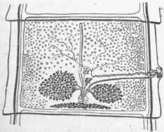
Fig. 174. - Unripe proglottis of T. solium showing sexual organs. The small vesicles scattered throughout are the male organs. The other structures shown are seminal tubes, vagina, globular body, yolk- body, ovaries, and unbranched uterus. x 10. (Lkuckart).
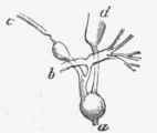
Fig. 175. - The globular body, or Mellis's body and its connections. See text. x30. (Leuckart).
The vagina forms a canal which passes transversely across the segment towards the middle line and tends also backwards, to end in a somewhat globular dilatation, sometimes called the Globular body or shell-gland, or Mellis's body. The connections of this body are difficult to make out, but they may be stated as follows, and understood by the annexed Figs. 174 and 175. In the posterior part of the segment, as shown in Fig. 174, are seen on either side the comparatively large ovaries, forming tree-like expansions, consisting of a congeries of closed tubes. The ovaries have ducts which pass into the globular body. Behind the ovaries and the globular body is the yolk gland, which is of a somewhat pyramidal shape and spread out laterally. This also communicates with the globular body in front of it. Besides these communications the globular body, which is thus the central part of the female organs, communicates with the uterus in front. At the period of development shown in figure the uterus consists of a simple tube extending longitudinally in the middle of the segment. It will thus be observed (Fig. 175) that the globular body has communication with four distinct structures, a with the yolk-sac, b with the ovaries, c with the vagina, and d with the uterus. The eggs pass from the ovaries first into the globular body, where they receive a covering of yolk, are fertilized, and undergo the beginning of their development. Then they pass into the uterus, which they fill up. As the ova accumulate in the uterus, this begins to throw out lateral branches to the number of seven to ten (see Fig. 176). The lateral branches often show considerable ramifications in this respect, and in their number contrasting with those of the next tape-worm. In the fully mature proglottis only the uterus crowded with ova is visible, the remaining organs having disappeared (see Fig. 173). The prominent ova often make the position and shape of the uterus very distinct, especially if the proglottis be spread out on a glass slide and allowed to dry.
Besides the sexual organs the proglottides possess muscular fibres, and a water-vascular system. The muscle is non-striated, and consists of longitudinal and transverse bundles. The water-vascular or excretory system (shown in Figs. 174 and 176) is in the form of tolerably wide channels, which begin at the head and are continued through the proglottides by two lateral channels right down to the last, where they open outwards. Near the posterior extremity of each proglottis the tubes form transverse communications (see figures). It is possible to inject these tubes from above downwards, but not from below upwards. In addition, the proglottides, as well as the head of the worm, possess numerous round or oval calcareous bodies, which are mainly in the superficial layers of the parenchyma.
Fig. 176. - Proglottis of T. solium, showing branching of uterus, x 5.
Continue to:
My Books

