1. Hematoma Of The Dura Mater. Pachymeningitis Hsemorrhagica
Description
This section is from the book "A Manual Of Pathology", by Joseph Coats, Lewis K. Sutherland. Also available from Amazon: A Manual Of Pathology.
1. Hematoma Of The Dura Mater. Pachymeningitis Hsemorrhagica
Haemorrhage on the internal surface of the dura mater, a lesion of considerable frequency, is found associated with thickening and new-formation, characters indicative of chronic inflammation. There are differences of opinion as to whether the inflammation is primary or secondary to the haemorrhage. The lesion presents itself mostly in the form of a somewhat massive blood-clot covering the internal surface of the dura mater and compressing the brain substance. When the clot is more particularly examined it is seen to be not exactly free on the surface of the dura mater, but covered with a delicate membrane, which is continued beyond the clot on the surface of the dura mater as a thin soft layer. This membrane generally has a brownish colour, evidently from the blood colouring-matter, and it presents in its substance, as well as between the membrane and the dura mater, numerous smaller haemorrhages. This, condition is of somewhat frequent occurrence, particularly among the insane.
As indicated above two views are held as to the nature of this condition, and it is quite possible that there may be actually two diseases. According to Prescott Hewett, Huguenin, and others, a haemorrhage into the cavity of the dura mater is the primary condition. It is undoubted that a haemorrhage may lead to a condition resembling that described. In a case of aneurysm of one of the larger cerebral vessels, where bleeding had occurred into the subdural space some time before the fatal cerebral haemorrhage, the author found a layer of soft tissue covering the dura mater and having much of the characters described above. In another case of the author's, an injury to the head was followed by a haemorrhage on the internal surface of the dura mater. The blood-clot was in great part replaced by an organized highly cellular membrane, in which were wide capillaries (see Fig. 344). This case had the further peculiarity (to be referred to further on) that the new-formed membrane became the seat of tuberculosis. In these and similar cases the coagulum on the surface of the dura mater becomes organized in the usual way, and vessels pass from the dura mater into the rudimentary tissue thus, produced. These thin-walled vessels (see figure) pecially apt to bleed for reasons to be presently referred to, and so there is haemorrhage in and under the new-formed tissue.
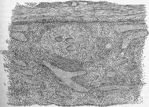
Fig. 344. - Dura mater with organized clot coating its internal surface. Above is dura mater. The new-formed tissue has wide capillaries, filled with blood-corpuscles. A tubercle with giant-cells in deeper part of new-formed tissue.
But many cases have a more spontaneous origin, and agree with the description which Virchow has given of a Pachymeningitis haemorrhagica. This disease begins in an inflammation of the dura mater, characterized by hyperaemia. The inflammation being chronic, the result is the formation of a soft membrane on the internal surface of the dura mater, owing to an inflammatory transformation of its internal layer. In its structure this membrane somewhat resembles mucous tissue, containing stellate and spindle-shaped cells in a matrix which gives a precipitate with acetic acid. In it there are large thin-walled blood-vessels in large numbers. The false membrane is easily lifted from the dura mater with forceps, and as this is being done numerous red threads are seen to stretch from it to the dura mater; these are the blood-vessels.
An explanation of the large size and tendency to rupture of these vessels has been suggested by Eindfleisch. To begin with, there is hyperemia of the dura mater with relaxation of the arteries. The normal capillaries of the dura mater being in a dense tissue are not readily dilated, but the blood in them is at a high pressure. The new-formed vessels, however, are delicate and lie in a soft tissue, and they communicate with the capillaries of the dura mater, in which the blood-pressure is excessive. They are therefore very liable to dilatation, and although they have the structure of capillaries they are mostly three or four times as wide as ordinary ones. These vessels often rupture, so that there is frequent haemorrhage into the soft membrane. But sometimes a more considerable haemorrhage occurs, and the blood accumulating dissects up the membrane from the dura mater, rupturing fresh vessels as it advances. In this way a large flat clot as thick as the hand may be formed, the proper haematoma. It will be observed that this clot is still covered with the membrane, and it is quite unusual to find the blood escaping into the cavity of the dura mater.
If a fatal haemorrhage does not occur, the new-formed membrane undergoes organization in the way of other inflammatory structures. It becomes more cellular and finally develops into connective tissue which coalesces with that of the dura mater. The disease, however, is apt to recur, arid a fresh soft layer is formed which goes through the same stages, so that there may be several layers in different stages of transformation on the surface of the dura mater, the innermost layer having the characters described above.
This condition occurs chiefly over the convexity of the brain, and is stated to be mainly in the domain of the middle meningeal artery. An acute suppurative inflammation very rarely develops in connection with the haematoma.
Continue to:
My Books

